ENDOSCOPIC ULTRASOUND(EUS)

EUS
Endoscopic ultrasound (EUS) is a minimally invasive procedure that combines endoscopy and ultrasound to obtain detailed images of the digestive tract and surrounding structures, such as the pancreas, liver, bile ducts, and lymph nodes. This advanced technique uses a flexible endoscope with a built-in ultrasound probe, which provides high-resolution images and allows for precise diagnosis and therapeutic interventions.
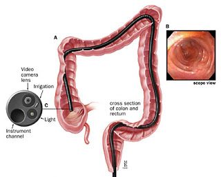
During the procedure, the endoscope is inserted through the mouth or rectum, depending on the area being examined. Ultrasound waves are then used to create detailed images of the walls of the gastrointestinal (GI) tract and nearby organs. EUS is particularly useful for diagnosing and staging cancers of the esophagus, stomach, pancreas, and rectum. It can also evaluate conditions such as chronic pancreatitis, cysts, gallstones, and bile duct abnormalities.
EUS is unique in its ability to perform fine-needle aspiration (FNA) or biopsy, where a small needle is guided into a suspicious area to collect tissue or fluid samples for laboratory analysis. This makes it a critical tool for identifying malignancies, infections, or other diseases without the need for open surgery.
Preparation for EUS is similar to other endoscopic procedures, involving fasting and possibly bowel preparation for rectal examinations. Sedation is typically used to ensure patient comfort. The procedure is generally safe, with a low risk of complications such as bleeding, infection, or perforation.
EUS offers superior imaging compared to external ultrasound or CT scans, allowing for more accurate diagnoses and targeted treatments. It has revolutionized the management of GI and hepatobiliary disorders by enabling early detection, precise staging, and minimally invasive therapeutic procedures. With its versatility and effectiveness, EUS has become an essential tool in modern gastroenterology and oncology care.
EUS is performed to:
- Evaluate tumors in the esophagus, stomach, rectum, pancreas, and bile ducts.
- Stage cancers (determine the extent of cancer spread).
- Obtain tissue samples (biopsies) of tumors or lymph nodes using fine-needle aspiration (FNA).
- Diagnose and evaluate pancreatic cysts.
- Evaluate bile duct stones or blockages.
- Guide interventions, such as celiac plexus block for pain management.
- Preparation typically involves:
- Fasting for several hours before the procedure.
- Adjusting or temporarily stopping certain medications as directed by your doctor.
- Discussing any allergies or medical conditions with your doctor.
- Fasting for several hours before the procedure.
- During the procedure:
- You will receive sedation to help you relax.
- An endoscope with an ultrasound probe is inserted through your mouth or rectum, depending on the area being examined.
- Ultrasound images are obtained to visualize the target area.
- If necessary, FNA is performed to obtain tissue samples.
EUS is generally not painful due to the sedation. Some discomfort or pressure may be felt.
The procedure typically takes 30 to 60 minutes, but it can take longer depending on the complexity of the case.

